Other Virus of Diarrhoeal Viruses Infection
Other_Possible_Enteric_Viruses
A. Caliciviruses (Sapoviruses)
Caliciviruses have been known to infect pigs, cats, sea-lions and fur-seals but it was not until 1976 that a calicivirus was fond in human faeces. The human calicivirus is morphologically indistinguishable from these animal strains. However, human calicivirus strains are antigenically distinct from animal strains. Human caliciviruses contain a single positive stranded RNA genome. and are about 33nm in diameter. The virus shows icosahedral symmetry, the face and the apices of the icosahedron are represented by cupped hollows, of which there are 32. This gives a virus a very characteristic appearance. However, not all the particles in the stool show the characteristic hollows and these particles can resemble Norwalk agents and SRSVs very closely. 4 serotypes of human calicivirus have been recognized, 3 in the UK, and 1 in Japan. No group antigen has been detected.
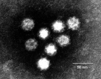
Electromicrograph of classical calicivirus particles. Note the charcteristic cupped shaped depressions on the surface
3 of the 4 human serotypes described are associated with outbreaks of diarrhoea and vomiting. Vomiting being a more prominent feature than diarrhoea and in this way and the epidemic nature of the viruses, infection by human caliciviruses resemble that by Norwalk agents. Caliciviruses have also been reported infrequently in the stools of normal babies. However it is not an easy virus to find and considerable under reporting may have taken place. Antibody against caliciviruses may be no more protective than with Norwalk, a further point of similarity with Norwalk agents. Volunteer studies in the UK and USA have shown the viruses to be capable of inducing disease in adult volunteers. The incubation period being 24 - 72 hours with symptoms of N+V, abdo pain and diarrhoea. Virus is excreted in the faeces during the illness. A serological survey carried out in Japan showed that the majority had acquired antibody in the first three years of life. The pattern of acquisition of antibody closely parallels the acquisition of antibody to rotaviruses and astroviruses, suggesting this virus is widespread although few actual infections are diagnosed. With much of the attention focused on noviruses, research on sapoviruses have been comparatively lacking. Recent studies that had been carried out with sensitive RT-PCR shows that like noroviruese, sappovirus infections are much more common than previously thought. Hopefully, more definitive date will become available in the near future.
B. Small Round Viruses
Small round virus-like objects are often seen in human faeces.
SRVs show consideration variation in size. Some of the smaller
SRVs have a diameter of 22nm and are often hexagonal in outline
so that they resemble parvoviruses. In some instances,
adenoviruses may also be found in the same stool and thus these
are probably defective adenovirus-associated virus. However,
adenoviruses are not always detectable and it may well be in
those instances that one is dealing with an autonomous
parvovirus. Indeed, certain animal parvoviruses e.g. canine
parvovirus, can cause severe diarrhoeal disease in animals. SRVs
in the next larger size band are about 25nm in diameter and
resemble enteroviruses. It is uncertain whether these are new
strains of enteroviruses but these viruses do not grow in cell
culture. Larger SRVs may occasionally be seen but it is doubtful
whether these are viruses. Occasionally, cubic bacteriophages may
be misdiagnosed as SRVs. SRVs may be present in a considerable
number of stools of infants. However, when detectable, the
numbers are relatively small. They are more likely to be found in
common source outbreaks. Both SRVs and SRSVs have been associated
with common source outbreaks, of which a considerable proportion
is associated with the consumption of shellfish, particularly
oysters. The incubation period is 36-48 hours, following which
the patient develops vomiting and diarrhoea. At present, there
are no known animal parallels.
C. Coronaviruses
Coronavirus-like particles are occasionally seen in stool extracts. The particles seen in the stools differ from the human respiratory coronaviruses in morphology. The particles in the stools have projections that resemble pins with a narrow shaft whereas those seen in the respiratory tract have club-shaped surface projections. The identities of these particles as true viruses have yet to be proved. Most attempts to culture coronaviruses have failed although there was a single unconfirmed report of growth in fetal intestinal cells. These particles have been seen in the faeces of adults as well as children. Prolonged excretion following recovery is common. Most reports associate these viruses with endemic rather than epidemic diarrhoea. No volunteer experiments have been reported to date. Coronaviruses are well established as causes of diarrhoea in animals, particularly in swine.
D. Other_Virus-like_Particles_Seen_in_Faeces
Breda-like Agents ;- Virus-like particles resembling those previously described in the faeces in cows have been seen in the faeces of a few children. Although an association with diarrhoea in man has been reported, further information is needed.
Bacteriophages ;- The human gut contains vast numbers of bacteria which are potential hosts for bacteriophages. Like animal viruses, bacteriophages come in a variety of sizes and shapes. However, the tailed bacteriophage has a structure unique to this class of virus. It is possible for cubic bacteriophages to be mistaken for SRVs. Moreover, a possible role for these phages in the causation of human diarrhoea should not be totally overlooked.