Hantaviruses
Interest in Hantaviruses has been stimulated by a marked increase in hantavirus infection being identified throughout Europe, and the discovery of hantavirus pulmonary syndrome. Significant progress has only been made in the last decade in elucidating the aetiology and the epidemiology of this disease, resulting in many different hantavirus isolates being obtained from human and rodent sources. Hantaviruses are responsible for causing a range of clinical manifestations collectively known as haemorrhagic fever with renal syndrome (HFRS) or, more recently hantavirus disease (HVD). Its first major impact on the Western world was during the Korean war, when over 3000 UN troops developed the disease with a 5-10% mortality rate. It was apparent that hantavirus disease was not new, and had probably been known by Russian, Chinese and Japanese for a number of years. The disease was recorded in Chinese medical records which were 1000 years old. The Japanese and Russian proved that the disease was caused by viruses when they carried out transmission experiments on prisoners of war. Records from previous wars showed that HVD was a severe problem, particularly associated with trench warfare e.g. American Civil War, WWI, Songo fever in the Japanese-Chinese war, "feldnephritis among German soldiers during 1941-42 in Russia and Finland. Despite extensive investigation during the Korean war and the period after, the breakthrough only occurred in 1978 when Lee et al. isolated the agent in Korean striped field mice. Serological examination of convalescent sera firmly associated the virus with the disease. A further breakthrough came in 1982 when sensitive cell cultures (Vero E6) became available for the isolation of the virus, and a large number of strains were isolated.
A. Properties
Member of the Bunyaviridae, enveloped ssRNA virus
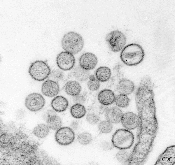
virion 98 nm in diameter, the surface has characteristic square grid-like structure.
Heat labile and is sensitive to lipid solvents
Genome consists of 3 -ve stranded segments; L, M, S.
At least 6 serotypes are known
Hantavirus particles (Source: CDC)
B. Pathogenesis and Clinical Features
The multisystem pathology of HVD is characterized by damage to capillaries and small vessel walls, resulting in vasodilation and congestion with hemorrhages. The damage is most severe during the hypotensive and the oliguric phases, when widespread haemorrhages occur. The kidney is usually involved resulting in a nephritis. Although immune complexes are found in the kidney, the amount is limited and it is thought that immunopathological mechanisms play a less important role in inducing nephritis than viral cellular destruction. Prior to serodiagnosis, the diagnosis of HVD was based on clinical judgment only. The severe form of the illness occurred in the Far East and is traditionally divided into 5 phases;
Febrile phase - after an incubation period of 2-3 days, the febrile phase begins abruptly, with fever, chills, headache, prostration, backache, anorexia, nausea and vomiting. There is a typical erythematous rash on the face, neck, shoulders and upper thorax.
Hypotensive phase - begins at fifth day of illness
Oliguric phase - begins at ninth day of illness. During the hypotensive and oliguric phase, the patient develops thrombocytopenia (100,000 platelet/mm2), proteinuria, and severe plasma loss through the damaged capillary epithelium. Moderate or severe acute renal failure may develop in severe cases. The worst cases go into thermal shock. Bleeding is usually confined to petechiae, and significant haemorrhage only occurs in 10% of cases. Death occurs in 5-10% of cases and usually result from shock and renal failure.
Diuretic phase - this occurs between days 12-14 and suggests improvement.
Convalescent phase - this may require up to 4 months.
The mild form of HVD seen in Scandinavia and occasionally
China is characterized by a sudden onset of fever, headache,
nausea, myalgia, lumbar and abdominal pain. This is a
self-limiting disease where nephritis leads to moderate renal
dysfunction and an uncomplicated recovery.
This newly defined group of viruses have been reported within many species of rodents and man worldwide. The HVDs reported in different parts of the world have now been confirmed as being caused by an antigenically-related group of viruses. These agents also share similarities in their epidemiological and ecological characteristics. Adult rodents show persistent infection without any clinical manifestations and secrete virus for prolonged periods. Following inoculation, a viraemia develops in the rodent during which, the virus is disseminated throughout the body. The virus is found in the lungs, spleen and kidneys for long periods in the rodent, perhaps for life. Saliva appears to play an important role in the horizontal transmission of the virus between rodents.
Man is probably infected through the respiratory route via aerosols of virus particles excreted by rodents in their lungs, saliva, urine and faeces. Transmission had also been documented following bites by rodents. Horizontal transmission among humans has not been documented, although blood and urine are infectious during th first 5 days of illness. Nosocomial spread was not documented during the Korean War. Although hantaviruses have been reported to have been recovered from some species of mites. It is generally accepted that arthropods are not necessary for transmission of the viruses between rodents and from rodents to man. Many hantavirus isolates have been isolated from humans and rodent hosts and are typed according to their serological cross- reactivity. There are at least 14 subtypes. The following are some of the subtypes associated with human disease.
Hantaan virus - found in China, Eastern USSR, carried by the stripped field mouse (Apodemus Agaris). Causes severe classical hantavirus disease.
Porrogia virus - found in Southern Europe, carried by the yellow-necked mouse (Apodemus flavicollis).Causes severe classical hantavirus disease.
Seoul type - have been identified in China, Japan, Western USSR, USA and S.America, carried by rats (rattus)
Puumala type - mainly found in Scandinavian countries, France, UK and the Western USSR. Carried by bank voles (Clethrionomys glareolus). Causes mild HVD (nephropathia epidemica)
Sin Nombre Virus - found in parts of the US, Canada, and Mexico. Carried by the deer mouse (Peromyscus maniculatus) and is associated with severe pulmonary disease: hantavirus pulmonary syndrome.
 |
 |
 |
 |
Rodent carriers of hantviruses: left to right - stripped field mouse, rat, bank vole, and deer mouse
The diagnosis of HVD is usually made on the clinical findings and serology results. In the early phase of the illness, the infection cannot be differentiated from other virus fevers. History should be sought for any possible contact with rodents.
Serological diagnosis - a variety of tests including IF, HAI, SRH, ELISAs have been developed
Direct detection of antigen - this appears to be more sensitive than serology tests in the early diagnosis of the disease. The virus antigen can be demonstrated in the blood or urine.
RT-PCR - this is now the test of choice for the early
diagnosis of hantavirus infection.
Virus isolation - isolation of the virus from urine is
successful early in the illness. Isolation of the virus
from the blood is less consistent.
Attentive and supportive therapy is important during the first
3 phases of the illness. Trials with ribavirin suggested a
reduction in mortality if the treatment is started within the
first 5 days of illness. A trial with a
-interferon was not successful. HVD is mainly a disease of
poverty and war. Control should be directed at reducing the
contact between rodent populations and humans. An inactivated
cell culture vaccine had been prepared in China where clinical
trials are due to begin soon. A portion of the Hantaan virus
genome has been cloned into vaccinia.
F. Comparative Clinical Features of Recognized
Hantavirus Disease_HVD
Nephropathia Far
Eastern
Rat-bourne Balkan
Epidermica
HVD
HVD
HVD
Virus type Puumala Hantaan Seoul Porogia
Overall Severity ..... 1-2 2-4 1-3 2-4
Multiphasic Disease occasionally Yes Blurred Yes
Hepatic abnormalities 0 0-1 1-3 0-1
Haemorrhagic phenomenon 0-1 1-4 1-2 1-4
Mortality <1% 5-10% 1% 5-35%
Score = 0 to 4
G. Hantavirus Pulmonary Syndrome
A unique hantavirus has been identified as the cause of the outbreak of respiratory illness (hantavirus pulmonary syndrome) first recognized in the "Four Corners" region SW USA in 1993. The rodent reservoir is the deer mouse. Onset of the illness is characterized by a prodrome consisting of fever, myalgia and variable respiratory symptoms eg. cough followed by the abrupt onset of acute respiratory distress. Headache and GI complaints have also been documented. Overall, the mortality rate had been 62%. The pathology is essentially the same as for the classical hantaviruses whereby damage to the capilliries and blood leakage occurs. In this case, the lungs rather than the kidneys are predominantly affected. Diagnosis is usually made by the demonstration of IgM by serological tests. RT-PCR and immunehistochemistry had also been reported to be useful. Virus isolation is not an option since the virus has never been isolated from clinical specimens.Ribavirin is being currently assessed in the treatment of hantavirus pulmonary syndrome. The virus involved is most closely related to the Prospect Hill virus and is called Sin Nombre virus. Deer mouse is distributed in most parts of the U.S., parts of Canada and Mexico. The risk to campers and hikers from hantaviruses is very small, the CDC have published guidelines for reducing the risk of exposure to rodents. Hantaviruses found in other rodents such as the white-footed mouse, the cotton rat and the rice rat have also been associated with hantavirus pulmonary syndrome. At present, at least three hantaviruses are associated with the syndrome.
Link to CDC's slide set on "Hantavirus Pulmonary Syndrome and Sin Nombre Virus"