Pathogenesis of Measles Virus Infections
D. Pathogenesis
Measles first gains access to the body via the upper respiratory tract or the conjunctiva. The virus quickly spreads to the immediate lymph nodes. Destruction of the lymphoid tissues leads to a profound leucopenia. A primary viraemia ensues which is responsible for spreading the virus throughout the rest of the R-E system and the respiratory system. A secondary viraemia follows whereby the virus is further spread to involve the skin, the viscera, kidney and bladder. The Koplik's spots and the rash in measles are thought to result from a delayed hypersensitivity reaction, the virus antigen being absent from the lesion itself.
Antibodies in the CSF are oligoclonal as opposed to the polyclonal response seen in the sera. This suggests that antibody in the CSF is made locally by a much smaller population of lymphocytes which have invaded this compartment. The M-protein is not recognized by the antibodies present in the CSF. SSPE brain lesions have M, N and P proteins present in infected cells whereas the envelope proteins are missing. The measles mRNAs isolated from SSPE patients showed a high rate of mutations, the highest rate of mutation in the M gene, followed by the F, H, P and N genes. In some cases, infectious MV particles may be recovered if the brain cells are co-cultured with tissue culture cells susceptible to measles virus. In other cases though, the block is only partially overcome and the agent remains cell associated. In this case, although MV envelope mRNAs are present, the envelope proteins are not synthesized. Another hallmark of SSPE is the hyperimmune response to measles antigens that include neutralizing antibodies in the serum and the CSF. In spite of this, the infection cannot be controlled. CMI is much more important than the humoral response in clearing measles virus infection. There is no evidence to suggest that the CMI is impaired in patients who develop SSPE.
Natural immunity to measles is known to last at least 65
years. In 1781 measles disappeared from the Faroe islands
following an epidemic and was not reintroduced until 1846.
Individuals old enough to have experienced the disease 65 years
previously were still protected. This unusual persistence of
immunity suggests that measles virus may normally persist inside
the body, possibly in lymphocytes so that immunity is
restimulated from within.
E. Diagnosis
The symptoms of acute measles are so distinctive that laboratory diagnosis is seldom required. However, as the vaccination program progresses, atypical forms of measles have emerged and laboratory diagnosis may be required.
Diagnosis of SSPE - the presence of measles specific antibodies in the CSF is the most reliable means of laboratory diagnosis of SSPE. Demonstration of MV-specific antibodies in the CSF may be sufficient with, if necessary, demonstration of MV-specific restricted heterogeneity by isoelectric focusing. Virus isolation from SSPE brain tissue is complicated. Alternately, brain biopsy material can be examined microscopically for inclusion bodies and virus antigen by immunofluorescence.
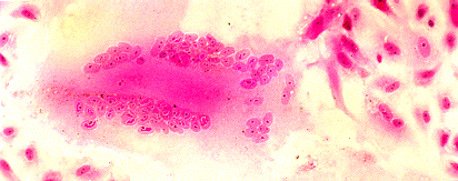
Syncytial formation caused by measles virus in cell culture (Courtesy of Linda Stannard, University of Capetown, S.A.)
F. Management
In the majority of patients, measles is an acute self-limiting disease that will run its course without the need for specific treatment. However, it is far more serious in the immunocompromised, the undernourished, and children with chronic debilitating diseases. Such patients can be protected by the administration of human anti-measles gammaglobulin if given within the first 3 days after exposure. Alternatively, the exposed individual can simply be vaccinated within 72 hours of exposure.
G. Prevention
With no animal reservoir, it must be possible to eradicate the virus through a controlled vaccination campaign. In the USA, where vaccination of all children is required before commencing school, case reports have fallen by over 99% but eradication has not been achieved. The following vaccines are available
The vaccination programme has been most effective in the USA, where measles immunization is compulsory. The incidence rate has also declined dramatically in the UK but without the rigorously pursued vaccination as practiced in the US, it is not likely to be as effective as that in N. America. In the third world, malnutrition aggravates measles infection and there are 900,000 measles related deaths per year. Vaccination in these areas has failed to yield dramatic results. The problem is that the vaccine is usually given at 12 months of age (it should not be given in younger individuals because the presence of maternal antibodies may lead to a poor response.) but infection in these areas often occurs earlier in life. Vaccination should therefore be performed on younger children than in the developed world. However, this must be balanced with the fact that the success rate is lower in younger children (50-75% in 6-month-old-children as opposed to 95% for 12-month-old children.). Measles is highly infectious and has a very high attack rate and thus it would be extremely difficult to eradicate the virus altogether through vaccination.
Management of Outbreaks
Measles outbreaks are most deleterious in wards with immunocompromised children or adults e.g. children with leukaemia and bone marrow transplant recipients. Measles is definitely as dangerous as VZV in that setting. HNIG should be given to all severely immunocompromised children irrespective of their immunization status since it has been reported that severe measles infection can occur in those who had been immunized and had a documented low-level antibody response. Therefore, the routine screening of children for measles antibody before admission is probably unjustified since there would be no difference in the management. The same argument applies to the screening of patients for immunity before the administration of HNIG. The use of live-attenuated vaccine for postexposure prophylaxis is contraindicated. The same protocol applies to immunocompromised adults who come into contact with measles. Immunocompetent children under 12 months in whom there is a particular reason to avoid measles, such as a recent severe illness, can also be given immunoglobulin. MMR vaccine should then be given after an interval of at least 3 months, at around the usual age.
![]()
![]()