Polyomaviruses constitute one genus of the family Papovaviridae.
The primate polyomavirus SV40 was discovered in 1960 as a
passenger virus in cultures of rhesus monkey cells. Poliovirus
vaccine produced in monkey kidney cells were contaminated by SV40
which was inadequately inactivated by formalin, and was
inadvertently administered to several million people. The 2 human
polyoma species, JC and BK, were isolated from patients with the
same initials in 1971. BK was isolated from the urine of a person
4 months after renal transplantation. JC was first isolated from
the brain from a patient suffering from Hodgkin's disease, and
suffering from PML. Previously, it had been shown that PML brain
tissue contained virus-like particles in the nuclei of abnormal
oligodendrocytes, which is the pathognomonic cell of the disease.
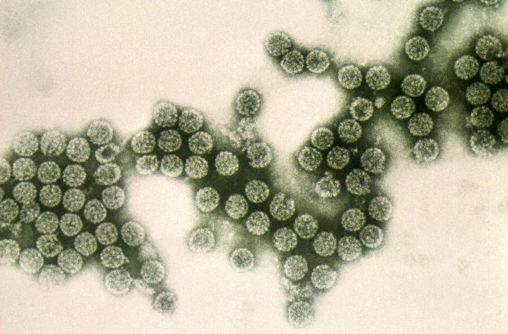
The appearance of these particles under EM strongly suggested
that they were polyomavirus particles. Strenuous attempts were
made to isolate the virus from 1965 onwards but success only came
in 1971 with the use of primary human glial cells. A lot of the
natural history of human polyomaviruses remain unknown, such as
the method of their transmission.
A. Properties
Belong to the family papovaviridae
naked dsDNA viruses with icosahedral symmetry
dsDNA molecule consists of around 5000 bp
5 transcripts are produced, both strands of DNA are used for
transcription. 5 proteins are produced
T and t are early proteins, VP1, VP2, and VP3 are the late
structural proteins.
The DNA of JC, BK and SV40 exhibit homology
T antigens of JC, BK and SV40 cross-react serologically and
functionally, although they are unique and distinct
JC and BK can agglutinate human and guinea pig RBCs
Able to transform certain cells in vitro, and to induce
tumors in experimental animals such as newborn Syrian
hamsters and adult owl monkeys.
Polyomaviurs virions
Polyomavirus replication in permissive cells can be detected
by observing CPE or plaque production, by identifying virus
particles in cell or culture fluid by EM, by detecting virus
antigens using IF, or by observing haemagglutination. JC will
only grow in a very restricted range of cells, mainly from the
brain. BK will grow in a wider range of cells including VERO. It
was demonstrated that BK is capable of supplying early gene
functions required by a temperature sensitive SV40 mutant for
growth at the non- permissive temperature, thus showing that the
T antigen of SV40 and BK can cross react functionally. Like many
other DNA viruses, JC and BK maintains a latent infection in the
body and are reactivated from time to time.
A number of other human polyomaviruses have been discovered
in recent years. Merkel Cell Polyomavirus was discovered in 2008 and is strongly
implicated in cellular transformation in an highly aggressive primary cutaneous
neuroendocrine skin neoplasm (associated with a poor prognosis) termed Merkel
cell carcinoma. KI polyomavirus was discovered by the Karolinska Institure in
2007 from respiratory specimens. It has yet to be associated with any disease.
WU polyomavirus (named for Washington University) was discovered in 2007 in a
nasopharyngeal aspirate from a three-year-old child with pneumonia. Like the KI
polyomvirus, it has been found in respiratory specimens worldwide but has yet to
be associated with any disease. Other recently discovered human polyomvirus
includes Human Polyomavirus 6 and Human Polyomavirus 7, Trichodysplasia
Spinulosa-Associated Polyomavirus, Human Polyomavirus 9 and Human Polyomavirus
10 (MW virus). The discovery of these new viruses suggests that polyomaviruses
may play a more significant role in human disease than previously understood.
JC and BK viruses are ubiquitous throughout the world, and the
2 viruses circulate independently. Isolated virgin populations
exist in remote areas of the world, BKV seems to have penetrated
more deeply into remote areas than JC. Antibody titres persist
throughout life. For JC, most persons become seropositive by the
age of 10, for BK, most persons are seropositive by 5. By
adulthood, 70-90% of individuals have antibodies to both JCV and
BKV. There is serological evidence for reactivation of JC and/or
BK in 5 to 10% of women during pregnancy, and virus can often be
isolated from the urine. Whether human intrauterine infection
with JC or BK occurs is still unresolved. Virus-specific IgM in
cord blood has been found by some workers but not by others.
Nevertheless, the possibility that congenital infections occur
cannot be excluded. In Germany, it was reported that 17 children
with various congenital disorders were found to have BKV-specific
IgM. There is no evidence for the existence of animal reservoirs.
The exact route of transmission is still unknown. By analogy with
murine polyoma virus and with SV40, infection may occur by
aerosol inhalation or oral ingestion of virus with excretion
occurring via the urinary tract.
Primary polyomavirus infections have not been associated with
any specific clinical syndromes. Most infections seem to be
subclinical although some children developed mild respiratory
symptoms and others had cystitis. It is thought that primary BKV
infections may on occasions be associated with either acute
respiratory disease or cystitis but further work is required.
Progressive multifocal leucoencephalopathy - JC
virus is now firmly associated with PML. It has not been
established whether PML is the result of a primary
infection with JCV in a person with impaired immunity or
whether it follows reactivation of latent virus. The fact
that PML is relatively uncommon in children and young
persons and more often develops in people in the fifth
and sixth decades of life suggests that latent virus is
the more likely cause. The pathogenesis of PML is not
fully understood but it is postulated that in patients
with disorders of immunoregulation, polyomaviruses are no
longer contained in a latent state and replicate within
the oligodendrocytes, causing the destruction of the cell
and the breakdown of the myelin sheath. PML is a unique
demyelinating disease which usually occurs in a person
with abnormal immune responses resulting from serious
disease, treatment with cytotoxic drugs or irradiation,
or long term immunosuppression. The pathology of PML is
distinctive and consists of multiple foci of
demyelination of varying size from pinpoint lesions to
areas of several centimetres. The lesions may occur
anywhere but are usually in the cerebral hemispheres,
less often in the cerebellum and brain stem and rarely in
the spinal cord. The oligodendrocytes in the peripheral
zone surrounding an area of demyelination are grossly
abnormal. The nuclei of abnormal oligodendrocytes are
packed with JC virions. Typically, PML evolves gradually,
with impairment of mental function and disturbance of
speech and vision. Movement may also be affected. The
disease then progresses rapidly and the patient is
severely disabled, eventually becoming demented, blind
and paralyzed and finally coma and death. PML is
frequently associated with lymphoproliferative and other
chronic diseases, such as AIDS, Hodgkin's disease, CLL,
sarcoidosis, TB, SLE and organ transplantation. Only
rarely has PML been reported occurring spontaneously in
an apparently healthy person. Occasionally, PML may
spontaneously arrest. PML has been reported in children
with congenital severe combined immunodeficiency which
suggests that a primary JCV infection is responsible.
Ureteric stenosis in renal transplant recipients -
the only other disease with which ureteric stenosis have
been associated is ureteric stenosis in renal transplant
patients. The polyomavirus infection induces
proliferation of the transitional epithelial cells in the
ureter and this can lead to partial obstruction or actual
stricture formation. The affected cells had inclusion
bodies. 9 cases have been recognized and both JC and BK
have been implicated. The ureteric obstruction occurred
between 50 to 300 days post-transplant.
Other possible associations - BK virus was
associated with cases of acute haemorrhagic cystitis
following bone marrow transplantation. However, it is
possible that two independent but synchronous events may
be taking place - reactivation of BKV and haemorrhagic
cystitis. The genomes of JC and BK virus were detected in
several tumors, but there is no evidence that human
polyomaviruses are associated with the causation of any
tumors.
PML - The clinical diagnosis of PML is confirmed
by histological and virological examination of brain
material obtained by brain biopsy or at postmortem.
Before a biopsy is done, both serum and CSF should be
examined for antibodies against JCV. A positive result
will not confirm PML, but a negative result makes the
diagnosis of PML very unlikely. It is rare to detect
antibodies against JC in the CSF. When it is detected, it
is suggestive of active multiplication of JCV within the
CNS. The brain biopsy or autopsy material can be examined
by EM or IEM. The specimen can also be examined directly
for JCV antigen by immunofluorescence or immunoperoxidase
staining and also by PCR. Virus isolation is very difficult for JCV. When
attempted, primary human fetal glial cells are used. The
presence of the virus in culture is confirmed by EM, IF
or haemagglutination. JC is rarely excreted in the urine
of patients suffering form PML.
Renal Tract Infections - the methods generally
employed to detect the presence of polyomavirus in urine
are cytological examination of the urine for
inclusion-bearing cells, EM and virus isolation. The
cytology of urine in human polyomavirus infection is
quite characteristic. The inclusion-bearing cells have a
characteristic appearance and are often present in large
numbers. Electron Microscopy of the urinary sediment
after centrifugation at 20000 may reveal the presence of
polyomavirus particles. It is difficult to isolate JC and
BK viruses: Primary PHFG cells must be used for isolation
of JCV; BKV have a wider host cell range and HEK cells
can be used as well as PHFG cells. More sensitive
techniques are being developed, such as dot- blot and
PCR.
Serological Diagnosis - HAI is the most widely
used serological technique for measuring antibodies
against the polyomaviruses. CFT, neutralization, ELISA
and RIAs have also been used.
Because of the invariably fatal outcome of PML, various
antiviral drugs have been tried including cidofovir and cytarabine. Despite
anecdotal reports of response to various treatments in the literature, all
controlled studies have failed to show any efficacy for the drugs tested against
PML One should also consider relaxing any immunosuppression regimes
in such patients.