Measles Virus
Measles is one of the typical viral diseases of childhood. However, unlike other common viral diseases i.e. VZV, rubella, mumps, and parvovirus infections, measles often leads to severe complications that may be fatal. In the third world, there may be up to 900,000 measles related deaths per year. Therefore, there is a lot of pressure on health in different countries in controlling the disease through vaccination. Indeed, measles is targeted by the WHO in its Expanded program of immunisation (EPI).
A. Properties
Member of the family of Paramyxoviruses
ssRNA enveloped virus, helical symmetry
lacks virion neuraminidase and thus grouped into a separate genus, the morbillivirus
Envelope consists of haemagglutinin protein and the fusion protein embedded in the lipid bilayer
M protein (membrane or matrix protein) lies immediately below the membrane.
ssRNA is encased in a helix of N (nucleocapsid protein). The ssRNA molecule is of negative sense.
The HA protein acts as a means of attachment to susceptible cells.
Measles is an antigenically stable virus. There is one serotype only and there are very little differences between different isolates.
The virus spreads by the respiratory route via aerosol
droplets and respiratory secretions which can remain infectious
for several hours. The infection is acquired through the upper
respiratory tract or conjunctiva. In the Northern Hemisphere, the
incidence tends to rise in the winter. In tropical regions
epidemics are less marked. In the prevaccination era, the maximum
incidence was seen in children aged 5 - 9 years. By the age of
20, approximately 99% of subjects have been exposed to the virus.
With the introduction of vaccine, measles infection has shifted
to the teens in countries with an efficient programme. In
contrast, in third world countries, measles infection has its
greatest incidence in children under 2 years of age. Here the
disease is a serious problem with a high mortality (10%) with
malnutrition being an important factor. In general measles
mortality is highest in children < 2 years and in adults. In
contrast to the influenza virus measles does not have an animal
reservoir.
After an incubation period of 10 - 11 days, the patient enters
the prodromal stage with fever, malaise, sneezing, rhinitis,
congestion, conjunctivitis and cough. Koplik's spots, which are
pathognomonic are measles, appear on the buccal and lower labial
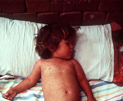
mucosa opposite the lower molars. The distinctive maculopapular
rash appears about 4 days after exposure and starts behind the
ears and on the forehead. From here the rash spreads to involve
the whole body. Cases of measles have been seen in partially
immunized children, in babies with residual antibodies, and in
people who have been given serum immune globulin for protection.
Occasionally, infection have also been seen in the course of live
vaccine failure. However, the symptomatology is very much
reduced.
Morbilliform rash of measles
Atypical measles infection may be seen in people who have been incompletely vaccinated. After an incubation period of about 7 - 14 days and is characterized by a sudden onset of high fever with headache, abdominal pain and myalgia. In contrast to acute measles, the rash develops on the distal extremities and spreads centripetally. The majority of cases develop a pneumonia. Occasionally, marked hepatosplenomegaly, hyperaesthesia, numbness or paraesthesia are also found.
Complications
Measles Pneumonia - this is a giant cell pneumonia
which occurs mainly in people with immunocompromised
patients. This is a severe infection with an often
protracted and fatal course. Measles infection is thus a
serious threat in immunocompromised and debillatated
patients.
Acute measles encephalitis - acute encephalitis is
a severe complication with a frequency of around 1 in
1000-5000. The mortality rate is around 15%, 20-40% are
left with residual neurological sequelae. Encephalitis
usually develops when exanthem is still present within a
period of 8 days after the onset of measles.
Occasionally, encephalitis may occur during the prodromal
stage. CSF findings in measles encephalitis consist
usually of mild pleocytosis and the absence of measles
antibodies.
Subacute measles encephalitis - This condition has
been recognized recently and only occurs in
immunosuppressed patients. It is most common in children
with leukaemia undergoing axial radiation therapy. The
incubation period ranges from 5 to 6 months. The
condition commences with focal convulsions, other signs
include hemiplegia, coma. This condition is frequently
confused with SSPE. However, the disease course is much
more rapid than SSPE and death supervenes within weeks or
a few months. No or only low titres of measles antibodies
are detectable in the CSF.
Subacute sclerosing panencephalitis (SSPE) - SSPE
is a rare slowly progressing fatal degeneration of the
brain. It is seen in children and young adults and occurs
6 - 8 years after the initial attack of measles. The
incidence is of the order of 1 in 100,000 cases of acute
measles. Half the SSPE patients have contracted measles
before the age of 2 years. The course of SSPE is highly
variable but usually starts with generalized intellectual
deterioration or psychological disturbance. It may be
several months before other neurological signs appear eg.
convulsions, aphasia, myoclonic jerks. In 75% of cases,
the retina, a chorioretinitis develops leading to
blindness. The progression of the disease is very
variable, the illness lasts from 1 to 3 years and
inevitably leads to death. Characteristic EEG changes are
present (Radermecker complexes) which are regarded by
some as pathognomonic for the disease. The CSF
characteristically has high levels of antibodies against
measles virus as well as elevated levels of
gammaglobulin. High levels of measles antibodies are also
present in the serum.
Myocarditis - myocardial deaths have been reported
during the prodrome and the acute phase of measles. ECG
abnormalities have been reported in up to 20% of children
with uncomplicated measles but frank measles
myopericarditis is rare.
Thrombocytopenic purpura - this is a rare
complication of measles and cases of DIC have
occasionally been reported.
Measles in pregnancy - measles in pregnancy result in a high rate of spontaneous abortion and premature delivery. There is some evidence that measles may be transmitted transplacentally as infants delivered during the mother's incubation often develops a rash simultaneously with the mother. While some infants with perinatally acquired measles have mild illnesses, others develop severe disease with pneumonia.
Measles first gains access to the body via the upper respiratory tract or the conjunctiva. The virus quickly spreads to the immediate lymph nodes. Destruction of the lymphoid tissues leads to a profound leucopenia. A primary viraemia ensues which is responsible for spreading the virus throughout the rest of the R-E system and the respiratory system. A secondary viraemia follows whereby the virus is further spread to involve the skin, the viscera, kidney and bladder. The Koplik's spots and the rash in measles are thought to result from a delayed hypersensitivity reaction, the virus antigen being absent from the lesion itself.
Acute measles panencephalitis - It is likely that
CNS involvement, even in uncomplicated measles, is
common. Transient EEG abnormalities are detected in 50%
of patients. Measles virus is rarely isolated from the
brain of a patient with acute measles panencephalitis.
Therefore, current theories favour an autoimmune reaction
as the possible cause of CNS damage.
Subacute measles encephalitis - arise only in
patients with severe immune disorder. Therefore it is not
usually accompanied by the formation of antibodies in the
CSF. Infectious virus has not been isolated by
conventional methods, suggesting defects in replication.
Recently biological studies on brain tissue from a case
of SME revealed that the envelope proteins were missing
from the brain tissue and only the N and the P protein
were consistently detected.
SSPE - in SSPE, the virus is first thought to gain entry to the CNS during the viraemia. Once there, it establishes a low-grade persistent infection. It is not known whether viral replication itself, or immunopathological mechanisms are responsible for the development of lesions. In SSPE, free infectious virus particles have never been isolated from the brain or the CSF, although some viral antigens may be found. Giant cells which are characteristic of acute measles infection are also absent. However, viral nucleocapsids are present in the cytoplasm. Therefore, some defect must exist in the virus replication process that prevents maturation. In the absence of free infectious particles, the infection may spread slowly by infectious nucleocapsids from cell to cell.
Antibodies in the CSF are oligoclonal as opposed to the polyclonal response seen in the sera. This suggests that antibody in the CSF is made locally by a much smaller population of lymphocytes which have invaded this compartment. The M-protein is not recognized by the antibodies present in the CSF. SSPE brain lesions have M, N and P proteins present in infected cells whereas the envelope proteins are missing. The measles mRNAs isolated from SSPE patients showed a high rate of mutations, the highest rate of mutation in the M gene, followed by the F, H, P and N genes. In some cases, infectious MV particles may be recovered if the brain cells are co-cultured with tissue culture cells susceptible to measles virus. In other cases though, the block is only partially overcome and the agent remains cell associated. In this case, although MV envelope mRNAs are present, the envelope proteins are not synthesized. Another hallmark of SSPE is the hyperimmune response to measles antigens that include neutralizing antibodies in the serum and the CSF. In spite of this, the infection cannot be controlled. CMI is much more important than the humoral response in clearing measles virus infection. There is no evidence to suggest that the CMI is impaired in patients who develop SSPE.
Natural immunity to measles is known to last at least 65
years. In 1781 measles disappeared from the Faroe islands
following an epidemic and was not reintroduced until 1846.
Individuals old enough to have experienced the disease 65 years
previously were still protected. This unusual persistence of
immunity suggests that measles virus may normally persist inside
the body, possibly in lymphocytes so that immunity is
restimulated from within.
The symptoms of acute measles are so distinctive that laboratory diagnosis is seldom required. However, as the vaccination program progresses, atypical forms of measles have emerged and laboratory diagnosis may be required.
Microscopy - production of multinucleate giant
cells with inclusion bodies is pathognomonic for measles.
During the prodrome phase, such cells are detectable in
the NPS (nasopharyngeal secretions). This is more rapid
and practical than virus isolation.
Immunofluorescence - direct and indirect
immunofluorescence have been used extensively to
demonstrate MV antigens in cells from NPS specimens. This
technique can also be applied to the urine as such cells
may be present in the urine 2 to 5 days after the
appearance of the rash. (Although like mumps, measles
virus is also excreted in the urine, this route is
unlikely to play a significant role in the spread of the
virus infection.)
RT-PCR - provides a sensitive and rapid means of
diagnosing measles infection.
Virus isolation - measles virus can be isolated
form a variety of sources, e.g. throat or conjunctival
washings, sputum, urinary sediment cells and lymphocytes.
Primary human kidney (HEK) cells are the best, although
primary monkey kidney can be used as well. Continuous
cell lines such as vero cells can also be used although
they are not as efficient as primary cell lines. A CPE
develops between 2 to 15 days, and consist of either a
broad syncytium or a stellate form with inclusion bodies
visible. The presence of measles can be confirmed by
haemadsorption. In acute measles, the isolation rate is
difficult and the success rate is low. Isolation is most
likely to be successful from material taken in the
prodrome phase but not in the later stages after the rash
has developed. Therefore isolation should only be
attempted in complicated cases such as suspected SSPE
where the lymphocytes may carry the virus, and in
immunocompromised individuals developing pneumonia.
4. Serology - diagnosis of measles infection can be made if the antibody titres rise by 4 fold between the acute and the convalescent phase or if measles-specific IgM is found. The methods that can be used include HAI, CF, neutralization and ELISA tests. Neutralization tests are the most sensitive but are not practical to perform. CFTs have a reduced sensitivity and thus are not useful for immune status screening.
Diagnosis of SSPE - the presence of measles specific antibodies in the CSF is the most reliable means of laboratory diagnosis of SSPE. Demonstration of MV-specific antibodies in the CSF may be sufficient with, if necessary, demonstration of MV-specific restricted heterogeneity by isoelectric focusing. Viral DNA may be detected in brain biopsy material by RT-PCR. Virus isolation from SSPE brain tissue is complicated. Alternately, brain biopsy material can be examined microscopically for inclusion bodies and virus antigen by immunofluorescence.
Syncytial formation caused by measles virus in cell culture (Courtesy of Linda Stannard, University of Capetown, S.A.)
In the majority of patients, measles is an acute self-limiting disease that will run its course without the need for specific treatment. However, it is far more serious in the immunocompromised, the undernourished, and children with chronic debilitating diseases. Such patients can be protected by the administration of human anti-measles gammaglobulin if given within the first 3 days after exposure. Alternatively, the exposed individual can simply be vaccinated within 72 hours of exposure.
Encephalitis - treatment of acute measles encephalitis is only symptomatic and supportive. A wide variety of treatment has been tried for SSPE but no convincing effects have been demonstrated.
With no animal reservoir, it must be possible to eradicate the virus through a controlled vaccination campaign. In the USA, where vaccination of all children is required before commencing school, case reports have fallen by over 99% but eradication has not been achieved. The following vaccines are available
Inactivated Vaccine - this vaccine was intended
for use in young children less than 1 year of age who are
most prone to severe complications. It was thought to be
advisable to avoid the use of a live vaccine. It was
found that at least 3 doses were needed to elicit a
protective antibody response but the antibody levels soon
waned. This leave the vaccinees open to attack by the
natural virus. In some cases, the nature of the partial
immunity led to serious hypersensitivity reactions to
infection (Atypical measles). The exact mechanism is
still uncertain but it was thought that the vaccine
lacked an important antigen of the virus and thus
immunity was not complete. In view of the above and the
fact that antibody levels decline rapidly after
administration of the killed vaccine, live vaccination is
now generally recommended and individuals previously
immunized with the killed vaccine should be reimmunized
with the live vaccine. The killed vaccine has now been
withdrawn.
Live vaccine - live vaccines are now usually used. The seroconversion rate is 95% and the immunity lasts for at least 10 years or more, possibly lifelong. The virulence of the attenuated strain now in use is so low that encephalitis has only been noted in 1 in 1 million recipients. SSPE has been reported in children given the live vaccine. However, the rate is lower than that following natural infection. Therefore the vaccine is safe for use in very young children. The live vaccine is now incorporated as part as the MMR vaccine. As vaccine-induced measles antibody develops more rapidly than following natural infection, MMR vaccine can be used to protect susceptible contacts during a measles outbreak. To be effective, the vaccine must be administered within three 3 days of exposure. If there is doubt about a child’s immunity, vaccine should be given since there are no ill effects from immunizing individuals who are already immune. Immunoglobulin should be given to those for whom the vaccine is contraindicated.
The vaccination programme has been most effective in the USA, where measles immunization is compulsory. The incidence rate has also declined dramatically in the UK but without the rigorously pursued vaccination as practiced in the US, it is not likely to be as effective as that in N. America. In the third world, malnutrition aggravates measles infection and there are 900,000 measles related deaths per year. Vaccination in these areas has failed to yield dramatic results. The problem is that the vaccine is usually given at 12 months of age (it should not be given in younger individuals because the presence of maternal antibodies may lead to a poor response.) but infection in these areas often occurs earlier in life. Vaccination should therefore be performed on younger children than in the developed world. However, this must be balanced with the fact that the success rate is lower in younger children (50-75% in 6-month-old-children as opposed to 95% for 12-month-old children.). Measles is highly infectious and has a very high attack rate and thus it would be extremely difficult to eradicate the virus altogether through vaccination.
Management of Outbreaks
Measles outbreaks are most deleterious in wards with immunocompromised children or adults e.g. children with leukaemia and bone marrow transplant recipients. Measles is definitely as dangerous as VZV in that setting. HNIG should be given to all severely immunocompromised children irrespective of their immunization status since it has been reported that severe measles infection can occur in those who had been immunized and had a documented low-level antibody response. Therefore, the routine screening of children for measles antibody before admission is probably unjustified since there would be no difference in the management. The same argument applies to the screening of patients for immunity before the administration of HNIG. The use of live-attenuated vaccine for postexposure prophylaxis is contraindicated. The same protocol applies to immunocompromised adults who come into contact with measles. Immunocompetent children under 12 months in whom there is a particular reason to avoid measles, such as a recent severe illness, can also be given immunoglobulin. MMR vaccine should then be given after an interval of at least 3 months, at around the usual age.
![]()
![]()